
Bladder And Bowel Management
Neurogenic bladder refers to the loss of normal bladder control due to a lesion in the central or peripheral nervous system. In spinal cord injury (SCI) the level and completeness of the injury determine whether the bladder behaves as an up…
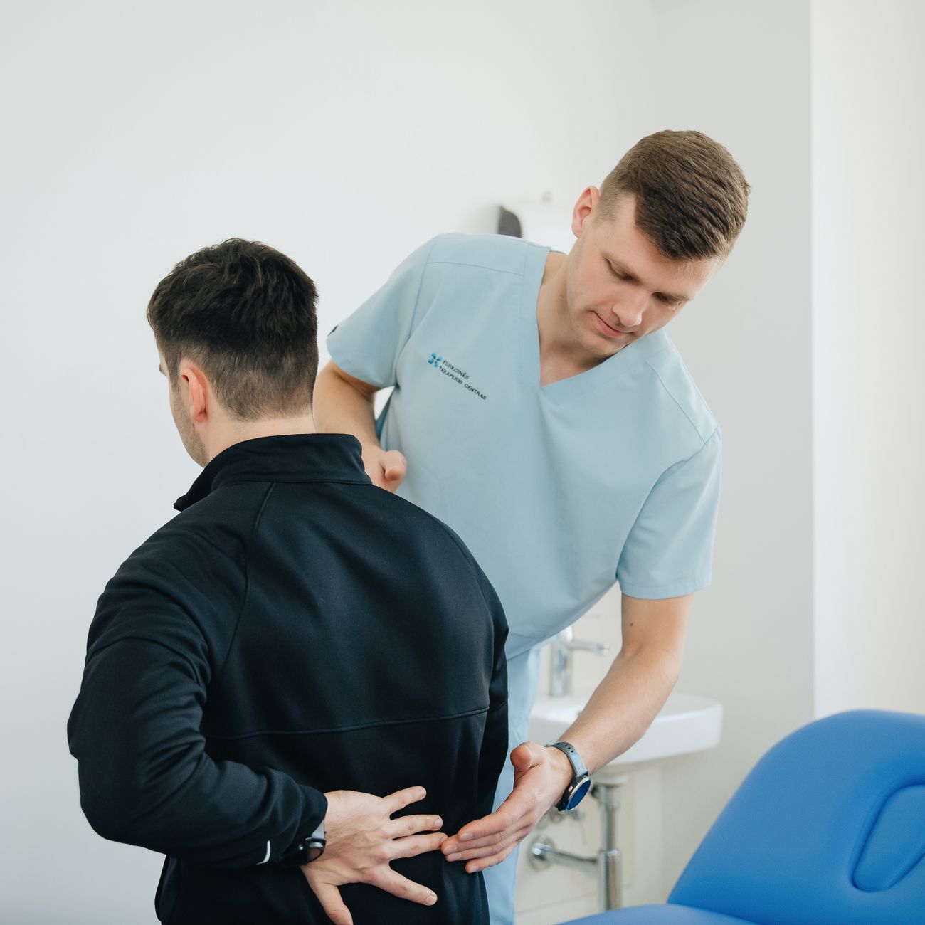
Neurogenic bladder refers to the loss of normal bladder control due to a lesion in the central or peripheral nervous system. In spinal cord injury (SCI) the level and completeness of the injury determine whether the bladder behaves as an upper motor neuron (spastic) or lower motor neuron (flaccid) system. Upper motor neuron lesions, typically seen in injuries above the conus medullaris, produce detrusor overactivity with an intact sphincter, whereas lower motor neuron lesions, such as conus or cauda equina injuries, result in detrusor areflexia and sphincter weakness. Understanding this distinction is essential for selecting appropriate management strategies, because the therapeutic goals differ: Inhibition of involuntary contractions in the spastic bladder versus facilitation of bladder emptying in the flaccid bladder.
Detrusor overactivity is a urodynamic finding characterized by involuntary detrusor contractions during the filling phase. These contractions may be spontaneous or triggered by stimuli such as a full bladder, coughing, or sudden movement. Clinically they manifest as urgency, frequency, and incontinence. Management options include anticholinergic medications (e.G., Oxybutynin, tolterodine), beta‑3 adrenergic agonists (e.G., Mirabegron), and intermittent catheterisation to maintain low bladder volumes and reduce the stimulus for overactivity. Patients with severe overactivity may benefit from intravesical botulinum toxin injections, which temporarily paralyze the detrusor muscle and increase bladder capacity.
Detrusor areflexia describes the absence of detrusor contractions during bladder filling, leading to urinary retention and high residual volumes. This condition is common after sacral or cauda equina injuries. The primary management goal is to ensure complete and regular emptying to prevent urinary tract infection (UTI) and upper urinary tract damage. Intermittent catheterisation, either clean or sterile, is the gold‑standard technique. In patients unable to perform self‑catheterisation, indwelling catheters (urethral or suprapubic) may be used, but they carry a higher risk of infection and urethral trauma. Bladder training, use of external collection devices for men, and scheduled voiding are adjunctive strategies.
Bladder compliance is the ability of the bladder wall to stretch without a significant rise in intravesical pressure. Low compliance indicates a stiff bladder, which can lead to high pressures that jeopardise renal function. Assessment of compliance is performed during urodynamic testing, where the change in volume divided by the change in pressure provides a numerical value (mL/cm H2O). Therapeutic measures to improve compliance include anticholinergic therapy, intravesical botulinum toxin, and, in refractory cases, surgical augmentation (e.G., Enterocystoplasty). Patients with low compliance are monitored closely with periodic renal ultrasound to detect hydronephrosis early.
Detrusor sphincter dyssynergia (DSD) occurs when the detrusor contracts against a closed external sphincter, creating high outlet resistance and potentially damaging the upper urinary tract. DSD is a hallmark of upper motor neuron bladder dysfunction and is identified on urodynamics by simultaneous pressure spikes in the detrusor and urethra. Management may involve intermittent catheterisation combined with timed voiding, pharmacologic agents such as alpha‑blockers to relax the sphincter, or electrical stimulation of the sacral roots (sacral neuromodulation). In severe cases, surgical sphincterotomy may be considered.
External urethral sphincter (EUS) is a striated muscle under voluntary control that contributes to urinary continence. In SCI patients, the EUS may become hypertonic (spastic) or hypotonic (flaccid) depending on the lesion level. Assessment of sphincter tone can be performed through digital rectal examination or electromyography (EMG). Pharmacologic agents such as tamsulosin (an alpha‑1 blocker) may reduce spastic tone, while pelvic floor muscle training can strengthen a weak sphincter. Understanding the functional status of the EUS guides decisions about catheter type and the need for adjunctive continence measures.
Urodynamic study (UDS) is an invasive diagnostic procedure that measures bladder pressure, capacity, compliance, and sphincter activity during filling and voiding phases. It is the cornerstone for classifying bladder dysfunction, guiding therapy, and monitoring disease progression. A typical UDS protocol includes cystometry, pressure‑flow study, and sometimes electromyography. The results help clinicians decide whether anticholinergic medication, intermittent catheterisation, or surgical intervention is indicated. Regular UDS, performed at least annually in high‑risk patients, can detect early changes that may predispose to renal damage.
Clean intermittent catheterisation (CIC) is a sterile‑technique catheterisation method where a catheter is inserted, the bladder emptied, and then removed. It is performed several times a day, typically every 4–6 hours, to maintain low bladder volumes. CIC reduces the risk of infection compared with indwelling catheters because it avoids a permanent foreign body. Training for CIC includes hand hygiene, catheter handling, and bladder volume monitoring. Patients must be educated on recognizing signs of infection, catheter blockage, and proper storage of catheters. For those unable to self‑catheterise, a caregiver can be trained to perform CIC under supervision.
Indwelling catheter (IC) refers to a catheter that remains in place for an extended period, either through the urethra (urethral catheter) or directly into the bladder via a suprapubic tract (suprapubic catheter). IC is indicated when CIC is not feasible due to hand function, severe spasticity, or patient preference. The main complications of IC are infection, urethral erosion, bladder stones, and bladder contracture. Preventive measures include regular catheter changes (every 2–4 weeks for urethral catheters, longer for suprapubic catheters), meticulous hygiene, and routine monitoring of urine characteristics. Suprapubic catheters are often preferred for long‑term use because they cause fewer urethral complications and are more comfortable for the patient.
Urethral catheter is a catheter inserted through the urethra into the bladder. It is the most common type of indwelling catheter but can cause urethral trauma, strictures, and discomfort. In males, the catheter is secured to the penis and leg; in females, it is taped to the thigh. The catheter size (French) is selected based on patient anatomy and the viscosity of urine. Smaller catheters reduce trauma but may increase blockage risk. Regular assessment of catheter integrity and securement is essential to prevent accidental dislodgement.
Suprapubic catheter is placed percutaneously through the lower abdomen into the bladder, usually under local anesthesia. It is indicated for long‑term bladder drainage, especially when urethral catheters are contraindicated. The insertion site is typically 2–3 cm above the pubic symphysis, and the catheter is secured with a retention disc. Suprapubic catheters allow for easier hygiene, less discomfort, and a lower rate of UTIs. However, they carry risks of bowel injury, catheter blockage, and skin infection around the insertion site. Patients should be taught to clean the site daily and to monitor for signs of leakage or infection.
Urinary tract infection (UTI) is the most common complication of neurogenic bladder. Symptoms may be atypical in SCI patients; fever, altered mental status, increased spasticity, or autonomic dysreflexia can be the only clues. Diagnosis relies on urine culture (>10⁴ CFU/mL) and clinical correlation. Management includes appropriate antimicrobial therapy based on sensitivity patterns, and preventive strategies such as regular bladder emptying, adequate hydration, and avoiding prolonged catheterisation. Prophylactic antibiotics are generally discouraged due to resistance concerns, but may be used in selected high‑risk individuals.
Autonomic dysreflexia (AD) is a life‑threatening condition that occurs in individuals with spinal cord lesions above T6. It is triggered by noxious stimuli below the level of injury, most commonly a distended bladder. The physiological response includes uncontrolled sympathetic discharge causing severe hypertension, bradycardia, headache, and sweating above the lesion level. Immediate management involves removing the stimulus, such as emptying the bladder, and monitoring blood pressure. Long‑term prevention includes maintaining bladder emptying schedules, using anticholinergic medication to reduce bladder overactivity, and ensuring catheters are not blocked.
Bladder capacity is the volume of urine the bladder can hold before the urge to void becomes overwhelming. Normal capacity ranges from 300 to 500 mL. In SCI patients, capacity may be reduced due to low compliance or increased due to chronic retention. Measuring capacity during cystometry provides guidance for catheterisation intervals; for example, a patient with a measured capacity of 250 mL may be scheduled to catheterise every 3–4 hours to keep residual volumes below 50 mL. Education on self‑monitoring bladder volume using a bladder scanner can empower patients to adhere to recommended intervals.
Residual urine volume (post‑void residual, PVR) is the amount of urine left in the bladder after voiding or catheterisation. High PVR (>100 mL) is associated with increased risk of infection and upper tract deterioration. In patients using CIC, PVR is measured after each catheterisation session using a portable bladder scanner. If PVR consistently exceeds the target, adjustments may be necessary: Increasing catheterisation frequency, using larger catheters, or reviewing anticholinergic dosing. Persistent high residuals may indicate the need for a urodynamic reassessment.
Hydronephrosis is the dilation of the renal pelvis and calyces due to obstructed urine flow. In SCI patients, it often results from high bladder pressures or DSD. Early detection is critical; routine renal ultrasound every 6–12 months can identify hydronephrosis before irreversible damage occurs. Management involves lowering bladder pressure through anticholinergics, intermittent catheterisation, or surgical interventions such as bladder augmentation or sphincterotomy. If left untreated, hydronephrosis can progress to renal insufficiency and eventual failure.
Urethral sphincter augmentation (e.G., Sphincterotomy, sphincteroplasty) is a surgical option for patients with refractory DSD or severe sphincter hypertonicity that cannot be managed medically. The goal is to reduce outlet resistance, thereby decreasing intravesical pressure and protecting the upper urinary tract. Sphincterotomy involves cutting the striated sphincter muscle, while sphincteroplasty reconstructs the sphincter to improve continence. Both procedures carry risks of incontinence and infection; therefore, they are reserved for carefully selected patients after exhaustive medical management has failed.
Bladder augmentation cystoplasty is a reconstructive surgery that increases bladder capacity and compliance by incorporating a segment of bowel (usually ileum or colon) into the bladder wall. This procedure is indicated for patients with low compliance, recurrent UTIs, and high intravesical pressures despite maximal medical therapy. Post‑operative care includes catheterisation training, monitoring for metabolic disturbances (e.G., Hyperchloremic metabolic acidosis), and regular surveillance for malignancy due to the presence of bowel tissue in the urinary tract. Long‑term outcomes show improved quality of life, but the surgery is complex and requires multidisciplinary follow‑up.
Botulinum toxin (Botox) injections into the detrusor muscle are an established minimally invasive treatment for refractory detrusor overactivity. The toxin blocks acetylcholine release at the neuromuscular junction, resulting in temporary paralysis of the detrusor. Injections are performed cystoscopically, typically delivering 200–300 units in 20–30 sites. Effects begin within 1–2 weeks and last 6–12 months. Patients must continue intermittent catheterisation during the effect period to avoid retention. Common side effects include urinary tract infection, haematuria, and occasional urinary retention requiring catheterisation.
Alpha‑adrenergic blockers such as tamsulosin or alfuzosin are used to relax the smooth muscle component of the urethral sphincter, thereby reducing outlet resistance. They are particularly helpful in patients with DSD who have a component of smooth muscle hypertonicity. These agents are taken orally once daily and have a favorable side‑effect profile, though they may cause hypotension or dizziness. Monitoring of blood pressure is recommended, especially in patients with autonomic dysfunction.
Anticholinergic medications block muscarinic receptors in the bladder, reducing involuntary detrusor contractions. Common agents include oxybutynin, tolterodine, solifenacin, and darifenacin. Dosing starts low and titrates upward based on efficacy and tolerability. Side effects such as dry mouth, constipation, and blurred vision are common; extended‑release formulations and transdermal patches can mitigate systemic effects. In patients with severe constipation due to bowel management, anticholinergics may exacerbate the problem, requiring careful balancing of bladder and bowel regimens.
Beta‑3 agonists like mirabegron stimulate beta‑3 adrenergic receptors in the detrusor muscle, promoting relaxation and increasing bladder capacity. They are an alternative or adjunct to anticholinergics, especially for patients who cannot tolerate anticholinergic side effects. Mirabegron is taken once daily and may increase blood pressure, so regular monitoring is advised. Combination therapy with an anticholinergic can provide synergistic benefits, but clinicians must be vigilant for additive side‑effects.
Pelvic floor muscle training (PFMT) is a rehabilitative approach that involves voluntary contraction and relaxation of the pelvic floor muscles to improve continence. In SCI patients with partial motor control, PFMT can be incorporated into a physiotherapy program to enhance sphincter strength. Biofeedback devices, such as surface EMG sensors, help patients visualise muscle activity and optimise training. PFMT is most effective when combined with other bladder management strategies and should be supervised by a trained therapist.
External collection devices (e.G., Condom catheters for men, female external urine collection systems) provide a non‑invasive method for urine drainage in patients who are unable to catheterise. They are best suited for patients with good skin integrity and no significant urethral pathology. Condom catheters must be sized correctly to prevent leakage or penile injury; they should be changed every 24–48 hours. Female external devices are less common due to anatomical challenges but may be used in select cases. These devices are not a substitute for regular bladder emptying via CIC or intermittent catheterisation, as they do not address detrusor pressures.
Bladder scanner is a portable ultrasound device that estimates bladder volume non‑invasively. It is an essential tool for home monitoring of bladder filling, allowing patients to schedule catheterisation before the bladder reaches unsafe volumes. Proper technique involves placing the probe suprapubically, aligning the image with the bladder axis, and interpreting the displayed volume. Training patients and caregivers in scanner use improves adherence to catheterisation schedules and reduces the risk of over‑distension.
Catheterisation technique includes hand hygiene, use of sterile gloves (for CIC), lubricating the catheter tip, gentle insertion to avoid urethral trauma, and complete drainage of the bladder before removal. For indwelling catheters, the technique also involves securing the catheter to prevent tugging, checking for patency, and documenting catheter change dates. Errors such as forceful insertion, failure to maintain a closed drainage system, or inadequate catheter size can lead to urethral injury, haematuria, and infection.
Spinal shock is the initial period after SCI during which reflexes are absent or diminished, and bladder function is typically atonic. This phase may last days to weeks, after which reflex activity returns, and the bladder may become spastic. Early assessment of bladder function should be delayed until the spinal shock phase resolves, because initial findings may be misleading. Once reflexes return, urodynamic testing can be performed to classify the bladder pattern accurately.
Upper urinary tract monitoring involves regular assessment of the kidneys and ureters to detect early signs of damage. Methods include renal ultrasound, serum creatinine measurement, and periodic urodynamic studies. In patients with high detrusor pressures (>40 cm H₂O), more frequent monitoring (every 3–6 months) is recommended. Any evidence of hydronephrosis, renal cortical thinning, or declining renal function prompts immediate intervention to lower bladder pressures.
Renal function tests include serum creatinine, estimated glomerular filtration rate (eGFR), and blood urea nitrogen (BUN). Baseline values are obtained shortly after injury and repeated at regular intervals. Persistent elevation of creatinine or reduction in eGFR signals possible upper tract compromise and necessitates a review of bladder management strategies. Adjustments may include intensifying catheterisation frequency, initiating anticholinergic therapy, or planning surgical decompression.
Continence devices such as absorbent pads, adult diapers, and penile clamps are adjuncts for patients with persistent incontinence despite optimal bladder management. These devices provide dignity and skin protection but do not address the underlying pathophysiology. Proper skin care, regular changing schedules, and education on signs of skin breakdown are essential components of continence device use. Over‑reliance on these devices without addressing bladder pressures can mask worsening urological conditions.
Bowel management terminology includes several key concepts that parallel bladder care. Neurogenic bowel describes the loss of voluntary control over bowel function due to spinal cord injury. Similar to bladder dysfunction, bowel dysfunction can be classified as upper motor neuron (spastic) or lower motor neuron (flaccid) based on the injury level. Upper motor neuron bowel presents with hyperreflexic sphincters, leading to constipation and potential fecal impaction. Lower motor neuron bowel results in loss of sphincter tone, causing incontinence and difficulty with evacuation.
Rectal tone is assessed by digital rectal examination and indicates the functional status of the internal sphincter. Hypertonic tone suggests spastic bowel, while hypotonic tone indicates flaccid bowel. Management strategies differ: Spastic bowel benefits from scheduled digital stimulation and laxatives, whereas flaccid bowel may require manual evacuation techniques and stool softeners.
Digital rectal stimulation (DRS) is a manual technique used to trigger the defecation reflex in patients with spastic bowel. The clinician or caregiver inserts a gloved finger into the rectum and applies rhythmic pressure to stimulate the recto‑anal inhibitory reflex, leading to relaxation of the external sphincter and facilitating stool passage. DRS is performed before a bowel routine, typically after a meal, to coordinate with the gastrocolic reflex.
Transanal irrigation (TAI) is a minimally invasive method where water is introduced into the colon via a catheter inserted into the rectum, flushing out stool. It is effective for both spastic and flaccid bowel patterns, reducing reliance on laxatives and decreasing the risk of impaction. TAI systems consist of a water container, a pump, and a rectal catheter. Training involves learning the correct volume of water (usually 500–1500 mL), insertion technique, and post‑irrigation care. Complications can include abdominal discomfort, rectal bleeding, and rare perforation; therefore, patients must be taught to recognize warning signs.
Laxatives are pharmacologic agents that increase stool frequency or soften stool consistency. They are classified as bulk‑forming agents (e.G., Psyllium), osmotic agents (e.G., Polyethylene glycol), stool softeners (e.G., Docusate sodium), and stimulant laxatives (e.G., Senna). In neurogenic bowel, osmotic agents are often preferred because they draw water into the colon, facilitating softer stools without excessive stimulation. Overuse of stimulant laxatives can lead to dependence and colonic dysmotility, so they should be used sparingly.
Stool softeners improve stool consistency by increasing water content, making evacuation easier. Docusate sodium is commonly used, but its efficacy alone is limited; it is most effective when combined with bulk‑forming agents and adequate fluid intake. Patients should be counseled to avoid excessive use, as softeners do not address the underlying neurologic dysfunction.
Fiber supplementation increases stool bulk, stimulating peristalsis. For SCI patients, soluble fiber (e.G., Oat bran) is generally better tolerated than insoluble fiber, which can cause gas and bloating. Fiber intake should be gradually increased to 20–30 g per day, accompanied by sufficient fluid intake (at least 1.5–2 L of water daily) to prevent constipation.
Fluid intake is a cornerstone of bowel management. Adequate hydration softens stool and promotes regular bowel movements. However, excessive fluid intake can increase urinary output and challenge bladder management, especially in patients with limited bladder capacity. Clinicians must balance fluid recommendations, often targeting 1.5–2 L per day, while tailoring to individual bladder and bowel needs.
Scheduled bowel program establishes a consistent routine for bowel evacuation, typically timed after meals to harness the gastrocolic reflex. The program may include a sequence of DRS, oral laxatives, TAI, and positioning strategies (e.G., Sitting on a commode with knees flexed). Consistency reduces variability in bowel habits, minimizes impaction risk, and improves patient confidence. Documentation of the program, including times, interventions, and stool characteristics, aids in evaluating effectiveness and making adjustments.
Positioning and assistive devices influence the effectiveness of bowel evacuation. Sitting upright with hips and knees flexed at approximately 90 degrees improves anorectal angle and facilitates defecation. Devices such as the “squatty potty” or a raised toilet seat can aid patients with limited trunk control. For those unable to sit, a commode with a supportive backrest and armrests can provide stability during DRS or TAI.
Abdominal massage is a technique that stimulates colonic motility. It involves gentle, clockwise strokes across the abdomen, starting at the right lower quadrant and moving upward toward the ribs, then across to the left side. Massage is performed before a bowel routine to encourage peristalsis, especially in patients with sluggish bowel activity. Contraindications include recent abdominal surgery, hernias, or severe abdominal pain.
Rectal suppositories contain stimulant agents (e.G., Bisacodyl) that act locally on the rectal mucosa to provoke a bowel movement. They are useful for quick evacuation when other measures have failed. Suppositories should be inserted after DRS or TAI, and the patient should be monitored for cramping or bleeding. Overuse can lead to mucosal irritation and reduced rectal sensitivity.
Enemas involve the instillation of fluid into the colon via a rectal tube to stimulate evacuation. Types include saline, mineral oil, and phosphate enemas. Enemas are effective for clearing impacted stool but must be used cautiously in patients with fragile rectal mucosa or those prone to perforation. Proper technique includes using a low‑pressure delivery system and limiting the volume to 500–800 mL.
Manual evacuation (ME) is a technique where the caregiver inserts a gloved finger into the rectum and gently removes stool. ME is indicated when stool is hard, impacted, or when other methods have failed. Caregivers must be trained in proper hand hygiene, lubrication, and gentle extraction to avoid mucosal injury. ME should be performed with the patient in a comfortable position, often lying on their side with knees drawn to the chest.
Colonic motility agents such as prucalopride, a selective serotonin 5‑HT4 receptor agonist, enhance colonic peristalsis. They are prescribed for patients with chronic constipation unresponsive to conventional laxatives. Side effects include headache and abdominal pain, and the medication should be initiated at a low dose with careful monitoring.
Pelvic floor electrical stimulation (PFES) uses low‑frequency electrical currents to activate the pelvic floor muscles, improving tone and coordination. PFES can be applied via surface electrodes placed on the perineum or via intramuscular probes for deeper stimulation. Sessions are typically 20–30 minutes, performed several times per week. PFES is particularly useful in patients with flaccid bowel who need to strengthen sphincter control.
Neurogenic bowel assessment includes a comprehensive history (frequency, consistency, incontinence episodes), physical examination (rectal tone, abdominal distension), and investigations (abdominal X‑ray, colon studies). Anorectal manometry may be performed to assess sphincter pressures and reflexes. The assessment guides the selection of interventions, from conservative measures to surgical options such as sacral anterior root stimulation.
Sacral anterior root stimulation (SARS) is an implanted neuroprosthetic system that delivers electrical impulses to the sacral nerve roots, synchronising bladder and bowel emptying with voluntary control. The device consists of a neurostimulator, electrode leads, and an external controller. When activated, SARS induces coordinated detrusor contraction and sphincter relaxation, enabling efficient voiding. It also stimulates colonic motility, aiding bowel evacuation. Candidate selection requires intact sacral reflex pathways, and the procedure carries surgical risks, including infection and hardware failure.
Colostomy is a surgical diversion of the colon to an abdominal stoma, used when bowel management cannot be achieved conservatively. Indications include severe fecal incontinence, chronic impaction refractory to all other measures, and high spinal lesions where sphincter control is impossible. A colostomy provides a reliable method for stool elimination, but it requires lifelong stoma care, skin protection, and psychological adaptation. Patients must be educated on appliance fitting, hygiene, and signs of stoma complications such as prolapse or stenosis.
Urethral sphincterotomy (also called sphincterotomy for bowel) involves cutting the external anal sphincter to reduce outlet resistance in patients with severe DSD. While this improves evacuation, it often leads to permanent incontinence, so it is reserved for cases where quality of life is severely compromised by constipation or impaction. The procedure is reversible only in limited circumstances, and patients must be prepared for the trade‑off between continence and ease of evacuation.
Quality of life measures in bladder and bowel management include validated questionnaires such as the Neurogenic Bladder Symptom Score (NBSS) and the Neurogenic Bowel Dysfunction (NBD) score. These tools assess symptom severity, impact on daily activities, and patient satisfaction with current management. Regular administration of these scales helps clinicians track progress, identify unmet needs, and adjust treatment plans accordingly.
Interdisciplinary care is essential for optimal bladder and bowel management. The team typically includes a physiatrist, urologist, gastroenterologist, rehabilitation nurse, occupational therapist, dietitian, and psychologist. Coordination ensures that interventions for bladder and bowel do not conflict (e.G., Anticholinergic medications worsening constipation) and that patient education is consistent across disciplines. Regular team meetings facilitate case review, goal setting, and shared decision‑making.
Patient education focuses on teaching self‑catheterisation techniques, recognizing signs of infection, understanding fluid and fiber intake, and adhering to scheduled bowel programs. Educational materials should be tailored to the individual's cognitive abilities, language preferences, and cultural background. Hands‑on demonstration, video tutorials, and written handouts improve retention. Reinforcement through follow‑up visits and home health visits enhances compliance.
Caregiver training mirrors patient education but emphasizes safe handling techniques, infection control, and emotional support. Caregivers must learn how to assist with CIC, manage indwelling catheters, perform DRS, and respond to emergencies such as autonomic dysreflexia. Providing caregivers with respite resources and support groups reduces burnout and improves overall care quality.
Complication monitoring includes routine urine cultures (especially after catheter changes), skin inspections around catheter sites, and assessment for signs of urinary retention (e.G., Increased spasticity, autonomic dysreflexia). Early identification of complications allows prompt intervention, preventing progression to more serious outcomes like renal failure or sepsis.
Technology integration offers innovative tools for bladder and bowel management. Mobile applications can remind patients of catheterisation times, track bladder volumes, and log bowel movements. Telehealth platforms enable remote urodynamic data review and medication adjustments. Wearable sensors that detect bladder fullness through acoustic or pressure signals are emerging, potentially reducing the need for frequent bladder scans.
Research directions continue to explore stem cell therapies for restoring neural pathways, gene therapy targeting muscarinic receptors, and advanced neuromodulation techniques such as closed‑loop sacral stimulation. Clinical trials investigating novel antimuscarinic agents with fewer systemic side effects are ongoing. Understanding the molecular mechanisms of neurogenic bladder and bowel will lead to more targeted, personalized treatments in the future.
Legal and ethical considerations involve informed consent for invasive procedures, especially when patients have limited decision‑making capacity. Documentation of patient preferences regarding catheter type, surgical interventions, and end‑of‑life care is vital. Confidentiality of urological and bowel health records must be maintained, and patients should be informed of their rights to privacy and autonomy in managing their conditions.
Insurance and reimbursement issues can affect access to essential equipment such as catheters, bladder scanners, and TAI systems. Clinicians should be familiar with coding for procedures like CIC training, urodynamic studies, and surgical interventions to facilitate coverage. Advocacy for policy changes that recognize the long‑term cost‑effectiveness of preventive bladder and bowel care is an important aspect of professional responsibility.
Psychosocial impact of bladder and bowel dysfunction is profound. Incontinence can lead to social isolation, depression, and reduced participation in community activities. Psychological support, peer support groups, and counseling services are integral components of a comprehensive rehabilitation program. Addressing body image concerns, especially after surgeries like suprapubic catheter placement or colostomy, improves overall wellbeing.
Transition to community living requires careful planning to ensure continuity of care. Home modifications may include installing grab bars in the bathroom, ensuring easy access to catheter supplies, and providing a private space for bowel routines. Home health nurses can assist with periodic catheter changes and monitoring for complications. Collaboration with community pharmacies ensures timely refill of medications and supplies.
Emergency management of bladder and bowel issues includes protocols for catheter blockage, acute urinary retention, severe constipation, and autonomic dysreflexia. Patients and caregivers should have a written emergency plan that outlines steps such as attempting to clear a blocked catheter, performing a rescue catheterisation, contacting emergency services, and administering antihypertensive medication if AD occurs. Regular drills reinforce confidence and speed of response.
Medication interactions are a key consideration. For example, anticholinergic agents used for bladder overactivity can exacerbate constipation; conversely, stimulant laxatives can increase bowel motility and potentially influence bladder emptying patterns. A thorough medication review should be conducted at each follow‑up visit, adjusting doses or substituting agents to minimise adverse effects while achieving therapeutic goals.
Nutrition and diet play a supportive role. A balanced diet rich in fruits, vegetables, and whole grains supplies necessary fiber for bowel health. Adequate protein intake supports tissue repair, especially after surgical interventions. Sodium restriction may be advised for patients with hypertension or renal impairment secondary to high bladder pressures. Hydration strategies should align with bladder capacity, avoiding large fluid loads that exceed safe bladder volumes.
Physical activity can improve both bladder and bowel function. Regular aerobic exercise enhances gastrointestinal motility and may reduce spasticity in the lower limbs, indirectly benefiting bladder control. Core strengthening exercises improve trunk stability, facilitating self‑catheterisation and bowel positioning. Physical therapists should design individualized programs that consider the patient’s level of injury, functional abilities, and fatigue thresholds.
Sleep hygiene influences bowel regularity. Disrupted sleep patterns can alter the circadian rhythm of colonic activity, leading to irregular bowel movements. Encouraging consistent sleep schedules, limiting caffeine intake, and addressing nocturnal bladder issues (e.G., Nocturnal enuresis) improve overall restorative sleep, which in turn supports bowel health.
Sexual function is intertwined with bladder and bowel management. Urinary incontinence can affect sexual activity, and catheter or stoma placement may cause discomfort during intercourse. Open communication with patients about concerns, providing options such as catheter‑friendly positions, and offering referrals to sexual health specialists are important aspects of holistic care.
Pregnancy considerations for women with SCI require specialized bladder and bowel management plans. Pregnancy increases intra‑abdominal pressure, potentially worsening reflux and urinary stasis. Adjustments may include more frequent catheterisation, temporary use of suprapubic catheters, and close monitoring for UTIs. Bowel regimens may need modification to avoid constipation that can lead to hemorrhoids or labor complications. Multidisciplinary collaboration with obstetrics, urology, and rehabilitation teams ensures safe maternal and fetal outcomes.
Long‑term follow‑up involves scheduled assessments of bladder and bowel status, renal function, skin integrity, and psychosocial health. Frequency of visits may be every 3–6 months for stable patients, or more often for those with recent changes in management or complications. Documentation of trends over time guides proactive adjustments, preventing deterioration and maintaining quality of life.
Case example 1 – A 28‑year‑old male with a T10 complete injury presents with frequent urgency, small‑volume voids, and occasional incontinence. Urodynamic testing reveals detrusor overactivity with low compliance and DSD. Management includes initiating oxybutynin XR, scheduling CIC every 3 hours, and adding tamsulosin to reduce sphincter tone. After three months, the patient reports fewer incontinence episodes, and bladder capacity increases to 350 mL with residual volumes <30 mL. Regular monitoring continues, with annual urodynamics to assess compliance.
Case example 2 – A 45‑year‑old female with a L1 incomplete injury experiences chronic constipation, occasional fecal incontinence, and occasional abdominal discomfort. Assessment shows hypertonic rectal tone and spastic bowel. A bowel program is instituted: High‑fiber diet (25 g/day), 2 L of water daily, scheduled DRS after each main meal, and polyethylene glycol 3350 at night. After six weeks, stool frequency improves to 1–2 soft stools per day, and incontinence episodes decrease. The patient is educated on using a portable bladder scanner to coordinate bladder emptying before bowel activities, minimizing the risk of autonomic dysreflexia.
Case example 3 – A 60‑year‑old man with a C5 AIS A injury uses a urethral indwelling catheter. He develops recurrent UTIs and presents with rising serum creatinine. Imaging shows mild hydronephrosis. After multidisciplinary review, the plan shifts to CIC with a suprapubic catheter for backup, combined with mirabegron to reduce detrusor overactivity. The patient receives training in CIC and is provided a portable bladder scanner. Six months later, UTIs decrease, renal function stabilises, and he reports increased independence.
Case example 4 – A 32‑year‑old woman with a T12 AIS B injury develops severe constipation unresponsive to oral laxatives. Colon X‑ray shows fecal loading. The team initiates TAI using a 1000 mL water load twice weekly, combined with a low‑dose stimulant laxative. After two months, bowel movements become regular, and the patient no longer requires manual evacuation. She reports improved confidence in social settings and reduced skin irritation from previous incontinence.
Case example 5 – A 55‑year‑old veteran with a C7 complete injury experiences high intravesical pressures (>50 cm H₂O) despite anticholinergic therapy and CIC. Urodynamics confirm low bladder compliance and DSD. The decision is made to proceed with sacral anterior root stimulation. Post‑operative testing shows coordinated detrusor contraction and sphincter relaxation, enabling efficient voiding with low residual volumes. The patient also reports improved bowel emptying due to enhanced colonic motility from the stimulation.
Practical tip 1 – Always document catheter size, type, and change date on the patient’s chart.
Key takeaways
- In spinal cord injury (SCI) the level and completeness of the injury determine whether the bladder behaves as an upper motor neuron (spastic) or lower motor neuron (flaccid) system.
- Patients with severe overactivity may benefit from intravesical botulinum toxin injections, which temporarily paralyze the detrusor muscle and increase bladder capacity.
- In patients unable to perform self‑catheterisation, indwelling catheters (urethral or suprapubic) may be used, but they carry a higher risk of infection and urethral trauma.
- Assessment of compliance is performed during urodynamic testing, where the change in volume divided by the change in pressure provides a numerical value (mL/cm H2O).
- Management may involve intermittent catheterisation combined with timed voiding, pharmacologic agents such as alpha‑blockers to relax the sphincter, or electrical stimulation of the sacral roots (sacral neuromodulation).
- Pharmacologic agents such as tamsulosin (an alpha‑1 blocker) may reduce spastic tone, while pelvic floor muscle training can strengthen a weak sphincter.
- Urodynamic study (UDS) is an invasive diagnostic procedure that measures bladder pressure, capacity, compliance, and sphincter activity during filling and voiding phases.